|
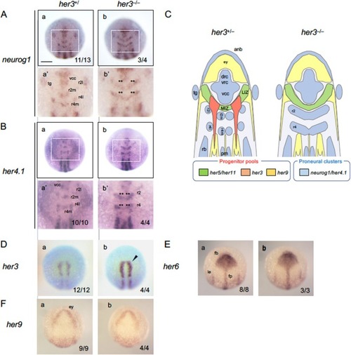
Phenotypic analysis of her3 mutant embryos. (A) Embryos from heterozygous mating (her3+/Δ2) were examined for the expression of neurogenesis‐related (A, B) and Notch‐independent her (D–G) genes by WISH, followed by genotyping. For each gene, boxed areas are enlarged below the whole views. Double asterisks (**) indicate ectopic expression in IPCD‐r2/IPCD‐4. Solid arrowheads show weak upregulation. Numbers of the embryos with the indicated expression patterns and total scored embryos are shown in the bottom‐right. her3+/+ and her3+/− embryos showed identical phenotypes and were grouped as her3+/, and scored accordingly; images show wild‐type (WT, her3+/+) embryos as representatives. Scale bars, 200 μm. (C) Schematic diagram, showing normal and ectopic neurogenesis observed in the IPCD in r1/2 (IPCD‐r2) and in r4 (IPCD‐r4) in WT embryos and her3 homozygotes, respectively. See the legends to Figures 1 and 3 for abbreviations.
|