
Fig. S4
- ID
- ZDB-FIG-241016-59
- Publication
- Lascaux et al., 2024 - TEX264 drives selective autophagy of DNA lesions to promote DNA repair and cell survival
- Other Figures
- All Figure Page
- Back to All Figure Page
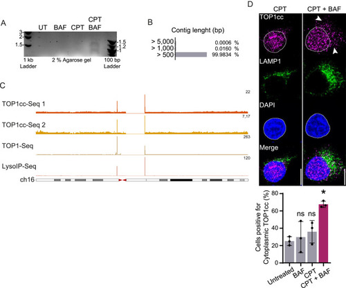
TOP1cc (TOP1 and its bound DNA fragment) are translocated in the cytoplasm and degraded by lysosomes, related to Figure 4 (A) DNA fragments purified by LysoIP after 6 h of treatment with 50 nM CPT and/or 50 nM BAF and run on a 2% agarose gel. UT, untreated. (B) Contigs length distribution was obtained by NGS sequencing of the DNA fragments purified by LysoIP after 6 h of treatment with CPT and 50 nM BAF. (C) Genomic browser images showing ChIP-seq of TOP1cc obtained in MCF7 (1) and LNCAP (2) (GEO: GSE135808),87 ChIP-seq of catalytically engaged TOP1 obtained in HCT116 (GEO: GSE57628),88 and LysoIP-seq signals obtained from sequencing lysosomal DNA after 50 nM CPT in HeLa. ChIP-seq and LysoIP profiles are presented at chromosome 16. All conditions were treated with CPT. Data aligned on genome hg38 and represented as sequence tags per million (TPM). (D) Immunofluorescence of TOP1cc foci and LAMP1 after 4 h of treatment with 50 nM CPT, with or without 50 nM BAF. Quantification of cells positive for cytoplasmic TOP1cc foci (n = 3). Scale bar, 10 μm. Two-way ANOVA. Error bar, SD. ∗p < 0.05; ns, not significant. |
Reprinted from Cell, 187(20), Lascaux, P., Hoslett, G., Tribble, S., Trugenberger, C., Antičević, I., Otten, C., Torrecilla, I., Koukouravas, S., Zhao, Y., Yang, H., Aljarbou, F., Ruggiano, A., Song, W., Peron, C., Deangeli, G., Domingo, E., Bancroft, J., Carrique, L., Johnson, E., Vendrell, I., Fischer, R., Ng, A.W.T., Ngeow, J., D'Angiolella, V., Raimundo, N., Maughan, T., Popović, M., Milošević, I., Ramadan, K., TEX264 drives selective autophagy of DNA lesions to promote DNA repair and cell survival, 5698-5718.e26, Copyright (2024) with permission from Elsevier. Full text @ Cell