
Fig. S3
- ID
- ZDB-FIG-241016-57
- Publication
- Lascaux et al., 2024 - TEX264 drives selective autophagy of DNA lesions to promote DNA repair and cell survival
- Other Figures
- All Figure Page
- Back to All Figure Page
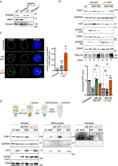
Autophagy promotes TOP1 degradation during replication stress to prevent protein aggregation, related to Figure 3 (A) Immunoblot showing TEX264 and RNF4 depletion in cells used for clonogenic assay. (B) Immunoblot of TOP1 level in total cell extract (n = 4). Treatment with 50 nM CPT was performed for 24 h, and 2 μM MG132 (MG) or 50 nM BAF was added in the last 8 h. Treatment with 1 μM CPT was performed for 3 h with MG132 or BAF. Quantification of normalized TOP1 protein level. One-way ANOVA, SEM represented; UT, untreated. (C) Immunofluorescence of γ-H2AX and 53BP1 foci after 1 h of 50 nM CPT or 1 μM CPT (n = 3). Scale bar, 10 μm. Quantification of positive nuclei for colocalized foci of both γ-H2AX and 53BP1. Two-way ANOVA. Error bar, SD. (D) Method for isolation of aggregated protein by sequential lysis and denaturation step. The aggregates fraction is insoluble in lysis buffer and 1.5% SDS but solubilized in 100% formic acid to be loaded on the gel. Isolation of the aggregates performed after 12 h of treatment with 50 nM CPT or 1 μM CPT (n = 3); UT, untreated. ∗p < 0.05; ns, not significant. |
Reprinted from Cell, 187(20), Lascaux, P., Hoslett, G., Tribble, S., Trugenberger, C., Antičević, I., Otten, C., Torrecilla, I., Koukouravas, S., Zhao, Y., Yang, H., Aljarbou, F., Ruggiano, A., Song, W., Peron, C., Deangeli, G., Domingo, E., Bancroft, J., Carrique, L., Johnson, E., Vendrell, I., Fischer, R., Ng, A.W.T., Ngeow, J., D'Angiolella, V., Raimundo, N., Maughan, T., Popović, M., Milošević, I., Ramadan, K., TEX264 drives selective autophagy of DNA lesions to promote DNA repair and cell survival, 5698-5718.e26, Copyright (2024) with permission from Elsevier. Full text @ Cell