|
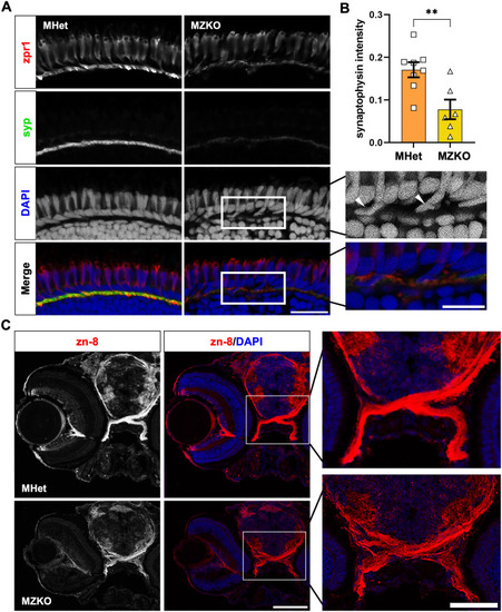
Retinal synapse formation and axon guidance deficits in MZKOs. A: Fluorescent staining of the outer layers of the retina showing comparable zpr1 (Arr3a) staining in the photoreceptor outer segments and pedicles in the outer plexiform layer, but reduced and disrupted synaptophysin (syp) staining, protrusion of photoreceptor cell bodies in the outer plexiform layer (arrowheads), and disorganization of horizontal cells lining the top of the outer nuclear layer (asterisks) suggesting defects to the ribbon synapses (40X magnification, scale bars: 20 µm for main panels, 10 µm insets). B: Quantification of synaptophysin intensity in the outer plexiform layer showing a significant reduction in MZKOs (**p = 0.0069). C: Fluorescent staining of transverse cryosections with zn-8 and DAPI showing an overall disruption in morphology of the retina with evidence of retinal ganglion cell axon defasciculation in the optic nerves at the chiasm (10X magnification, scale bars: 100 µm for main panels, 20 µm for chiasm insets).
|