|
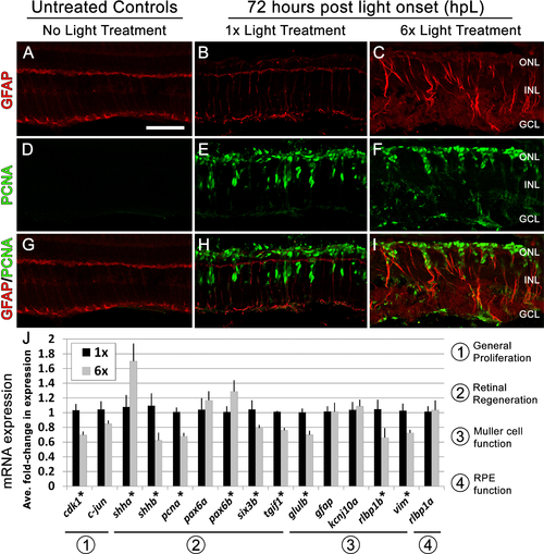
Following six rounds of light treatment Müller glia remain persistently gliotic at the canonical peak stage of progenitor proliferation and migration.(A–I) Retinal sections collected at 72 hpL immunolabeled with anti-GFAP (red) and anti-PCNA (green) in untreated control (No Light Treatment), experimental control (1×Light Treatment), and experimental retinas (1×Light Treatment). (A–C) Müller glia are immunolabeled with anti-GFAP. (D–F) Müller glia that have re-entered the cell cycle are immunolabeled with anti-PCNA. (G–I) Merge of anti-GFAP and anti-PCNA immunolabeling. (J) Graph showing the average fold-change in expression of genes associated with (1) general proliferation (cdk1, c-jun), (2) retinal regeneration (shha, shhb, pcna, pax6a, pax6b, six3b and tgif1), (3) Müller cell function (glulb, gfap, kcnj10a, rlbp1b, vim), and (4) RPE function (rlbp1a). Asterisk indicates significantly different from control (p < 0.05). Scale bar represents 25 μm.
|