FIGURE SUMMARY
Title

Gdf11 regulates left-right asymmetry development through TGF-β signal

Authors
Yao, W., Wei, Z., Tian, X., Tan, J., Liu, J.
Source
Full text @ Cell Prolif.

gdf11 expression pattern and mutant construction. (A) Whole‐mount in situ hybridization with gdf11 from the 2‐cell stage to 72 hpf. gdf11 sense probe was utilized as negative control at 1 cell stage. (B) Schematic picture showing the wild type and mutated site of gdf11−/− mutants. (C) Western blots of lysates from WT and gdf11−/− embryos using antibodies against Gdf11 with β‐tubulin as a loading control. (D) Alcian blue staining in both WT and gdf11 mutants. gdf11 mutants exhibit defects in the alignment of upper and jaw elements, the angle of ch. articulation, and the morphology of the op. The defects of the alignment of upper/jaw elements and the morphology of the op in gdf11 mutant were marked with straight lines and arrows in the picture. op,opercle; ch,ceratohyal. Scale bar, 50 μm. (E) Live images of WT and gdf11−/− mutants from 3 to 5 dpf. Black triangle indicates craniofacial cartilage. Scale bar, 100 μm.

gdf11 is essential for left–right patterning. (A) Cardiac looping and liver/pancreas positions analysed at 48 hpf. Heart looping visualized by in situ hybridization with cmlc2 probe at 48 hpf (top panel). A, atrium; V, ventricle. Liver/pancreas positions visualized by in situ hybridization with hhex probe at 48 hpf (bottom panel). L, liver; P, pancreas. (B) Graph showing the percentage of embryos with normal D‐looping (dark blue), no looping (yellow), or L‐looping (green). (C) Graph showing the percentage of embryos with normal liver/pancreas position (dark blue), bilateral liver/pancreas position (yellow), or reverse liver/pancreas position (green). (D) spaw (upper panel) and pitx2 (lower panel) expression in WT and gdf11−/− mutants at 21 or 22 ss. (E, F) Percentage of spaw (E) and pitx2 (F) expression in WT and gdf11−/− mutants. Normal (dark blue), reverse (yellow), or absent (green). (G, H) Graphs depicting the expression of midline ntl at 19 ss (G) and the percentage of ntl expression in WT and gdf11−/− mutants (H). (I, J) Graphs depicting the expression of lft1 at 16 ss (I) and the percentage of lft1 expression in WT and gdf11−/− mutants (J).

gdf11 is required for DFC proliferation and KV cilia formation. (A) Live image of Kupffer's vesicle at the 10‐somite stage in both WT and gdf11−/− mutants. Scale bar, 200 μm. (B) Percentage of KV size in WT and gdf1−/− mutants. (C) in situ hybridizations of charon were used to assess Kupffer's vesicle lumen size at 6 ss. Scale bar, 100 μm. (D) Visualization of KV cilia at the 10‐somite stage using an anti‐acetylated tubulin antibody in control or gdf11−/− embryos of the Tg(sox17‐GFP) line, showing both KV cells (green) and cilia (red). Scale bar, 50 μm. (E,F) Number (E) and length (F) of KV cilia in WT and mutant embryos. Graph indicates the mean of cilia number and length, error bars represent the standard deviation. Statistical significance between WT and gdf11−/− mutants was determined using a two‐tailed unpaired t‐test. (G, H) Confocal images of 75% epiboly zebrafish embryos with pH3 staining in a Tg(sox17‐GFP) background (G). Quantification of pH3 positive cells (H). Error bars represent the standard deviation. Statistical significance was determined using a two‐tailed unpaired t‐test. Scale bar, 50 μm. (I, J) Confocal images of 75% epiboly zebrafish embryos with TUNEL staining in a Tg(sox17‐GFP) background (I). Quantification of TUNEL positive cells (J). Error bars represent the standard deviation. Statistical significance was determined using a two‐tailed unpaired t‐test. Scale bar, 50 μm.

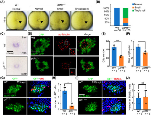
Gdf11 forms heterodimer with Spaw and regulates DFC specification through TGF‐β signal. (A) Immunoprecipitation between zGdf11‐Flag and zSpaw‐Myc in HEK 293T cells. (B) Western blot results of overexpressed zGdf11‐Flag and zSpaw‐Myc in HEK 293T cells with or without SB431542 treatment. (C) foxj1a and its target gene expression using WISH in WT or gdf11 mutants with or without treatment. (D–G) Expression levels of foxj1a and its target genes. Error bars represent the standard deviation. Statistical significance was determined using a two‐tailed unpaired t‐test. (H) Visualization of KV cilia at the 10‐somite stage using an anti‐acetylated tubulin antibody in both WT or gdf11−/− mutants with or without SB431542/SRI‐011381 treatment. Scale bar, 50 μm. (I, J) Number (I) and length (J) of KV cilia in WT and mutant embryos with or without SB431542/SRI‐011381 treatment. Graph indicates the mean of cilia number and length, error bars represent the standard deviation.

Mechanism diagram of Gdf11 regulating left–right asymmetry development. Gdf11 forms a heterodimer with Spaw, activating the activin receptor and phosphorylating Smad2/3. This process DFC proliferation, subsequently activates the expression of foxj1a and its target genes, promoting cilia formation. The diagram was drawn using Adobe Illustrator 2020 software.

Acknowledgments
This image is the copyrighted work of the attributed author or publisher, and ZFIN has permission only to display this image to its users. Additional permissions should be obtained from the applicable author or publisher of the image. Full text @ Cell Prolif.