
- Title
-
Successful large gene augmentation of USH2A with non-viral episomal vectors
- Authors
- Toms, M., Toualbi, L., Almeida, P.V., Harbottle, R., Moosajee, M.
- Source
- Full text @ Mol. Ther.
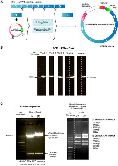
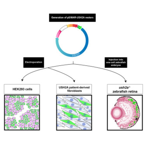
Generation of pS/MAR-USH2A vectors (A) pS/MAR-USH2A vectors were generated by inserting the full USH2A coding sequence (16 kb) into the backbone in 5 fragments. The vectors contained a CAG or CMV promoter and P. plumata GFP sequence 5′ of the USH2A sequence and an S/MAR sequence at the 3′ end. The P2A sequence encodes a self-cleaving peptide. (B) PCR was performed to amplify each USH2A fragment. (C) pS/MAR backbone digestion cut out the puromycin cassette and restriction enzyme digestion controls of the pS/MAR-USH2A vectors using SAPI. |
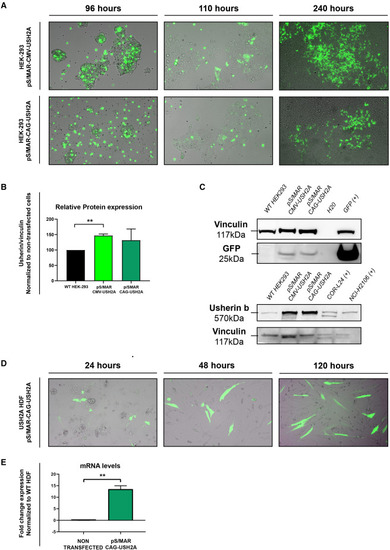
Transfection of HEK293 cells and USH2A patient-derived human dermal fibroblasts (A) Fluorescence microscopy images showing GFP expression in HEK293 cells electroporated with pS/MAR-CMV-USH2A and S/MAR-CAG-USH2A vectors at 96, 110, and 240 h post transfection. (B and C) Western blot analysis of usherin expression was performed in non-transfected HEK293 cells and pS/MAR-CMV-USH2A- and pS/MAR-CAG-USH2A-transfected HEK293 cells. GFP was also detected, and vinculin was used as a control. Two cancer lines, COR-L24 and NCI-H2106, were used as positive controls for the usherin protein band. Full blot images for usherin bands are available in the supplemental information. (D) Fluorescence microscopy images showing GFP expression in USH2A patient-derived human dermal fibroblasts electroporated with the pS/MAR-CAG-USH2A vector at 24, 48, and 120 h post transfection. (E) Fold change gene expression of USH2A in non-transfected or pS/MAR-CAG-USH2A-transfected USH2A patient fibroblasts, normalized to non-transfected WT fibroblasts. ∗∗p < 0.01. |
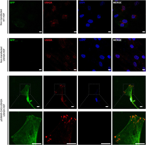
Over-expression of usherin in dermal fibroblasts transfected with pS/MAR-USH2A USH2A patient-derived dermal fibroblasts, non-transfected or transfected with pS/MAR-CAG-USH2A, were immunostained with anti-usherin (red). GFP (green) was also detected, and DAPI nuclei stain (blue) was used. Scale bars, 10 μm. |
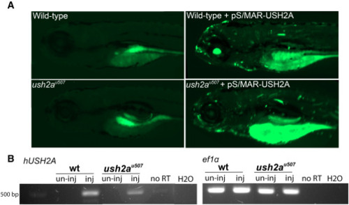
pS/MAR-USH2A vector expression in zebrafish (A) WT and ush2au507 zebrafish were micro-injected with pS/MAR-CMV-USH2A at the one-cell stage. GFP expression was detected in injected WT and ush2au507 larvae at 5 dpf. (B) RT-PCR analysis detected human USH2A (hUSH2A) expression in injected (inj) zebrafish and not in un-injected (un-inj) larvae. ef1a gene expression was used as a control. No reverse transcription (no RT) and water (H2O) controls were performed. The full gel is available in the supplemental information. |
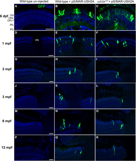
Long-term expression of pS/MAR-USH2A in the zebrafish retina GFP expression was observed in the retina of WT and ush2au507 zebrafish injected with pS/MAR-CMV-USH2A at the one-cell stage. Anti-turboGFP was used to aid GFP detection on retinal sections at ages 6 days post-fertilization (dpf) (A–C), 1 month post-fertilization (mpf) (D–F), 2 mpf (G–I), 3 mpf (J–L), 6 mpf (M–O), and 12 mpf (P–R). Sections were counterstained with DAPI nucleic acid stain (blue). OS, photoreceptor outer segment; ONL, outer nuclear layer; OPL, outer plexiform layer; INL, inner nuclear layer; IPL, inner plexiform layer; ON, optic nerve. Scale bars, 25 μm. |
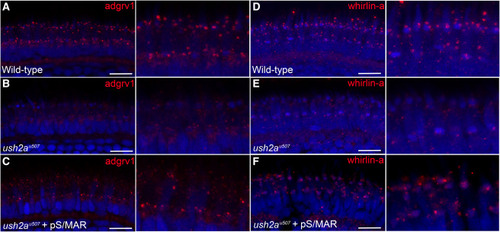
Partial rescue of the Usher 2 protein complex in pS/MAR-USH2A-injected ush2au507 zebrafish Retinal sections from zebrafish at 6 dpf were immunostained with antibodies for two Usher 2 complex proteins: adgrv1 (A–C) and whirlin-a (D–F). Specific punctate expression of adgrv1 (red, A) and whirlin-a (red, D) was detected in the WT photoreceptors, while both proteins were mislocalized in the ush2au507 retina (B and E). In ush2au507 zebrafish injected with pS/MAR-CMV-USH2A at the one-cell stage, specific adgrv1 and whirlin-a expression could be observed in some photoreceptors (C and F). Scale bar, 10 μm. |
|