
Image
|
Figure Caption
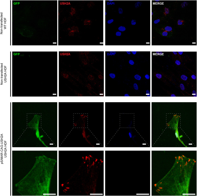
Fig. 3 Over-expression of usherin in dermal fibroblasts transfected with pS/MAR-USH2A USH2A patient-derived dermal fibroblasts, non-transfected or transfected with pS/MAR-CAG-USH2A, were immunostained with anti-usherin (red). GFP (green) was also detected, and DAPI nuclei stain (blue) was used. Scale bars, 10 μm.
Acknowledgments
This image is the copyrighted work of the attributed author or publisher, and
ZFIN has permission only to display this image to its users.
Additional permissions should be obtained from the applicable author or publisher of the image.
Full text @ Mol. Ther.