
Fig. 2
- ID
- ZDB-FIG-231221-44
- Publication
- Toms et al., 2023 - Successful large gene augmentation of USH2A with non-viral episomal vectors
- Other Figures
- All Figure Page
- Back to All Figure Page
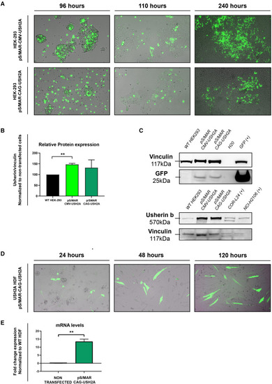
Transfection of HEK293 cells and USH2A patient-derived human dermal fibroblasts (A) Fluorescence microscopy images showing GFP expression in HEK293 cells electroporated with pS/MAR-CMV-USH2A and S/MAR-CAG-USH2A vectors at 96, 110, and 240 h post transfection. (B and C) Western blot analysis of usherin expression was performed in non-transfected HEK293 cells and pS/MAR-CMV-USH2A- and pS/MAR-CAG-USH2A-transfected HEK293 cells. GFP was also detected, and vinculin was used as a control. Two cancer lines, COR-L24 and NCI-H2106, were used as positive controls for the usherin protein band. Full blot images for usherin bands are available in the supplemental information. (D) Fluorescence microscopy images showing GFP expression in USH2A patient-derived human dermal fibroblasts electroporated with the pS/MAR-CAG-USH2A vector at 24, 48, and 120 h post transfection. (E) Fold change gene expression of USH2A in non-transfected or pS/MAR-CAG-USH2A-transfected USH2A patient fibroblasts, normalized to non-transfected WT fibroblasts. ∗∗p < 0.01. |