|
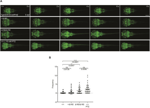
slc25a22aΔ182/Δ182 larvae exhibit elevated Ca2+ propagation from the midbrain to the spinal cord. (A) Tg(elavl3:Gal4,UAS:GCaMP6s) embryos at 5 dpf expressing GCaMP6s, a Ca2+ indicator, in mature neurons were subjected to time-lapse imaging for 20 min. WT larvae were exposed to 10 mM pentylenetetrazol (PTZ) for 30 min before the imaging. Scale bar: 300 μm. (B) Frequency of Ca2+ propagation from the midbrain to the spinal cord was measured for 20 min. Data are presented as mean±s.d. ns, not significant; **P<0.01 and ****P<0.0001 by one-way ANOVA with Tukey's HSD post hoc test (slc25a22a+/+, n=39; slc25a22a+/Δ182, n=40; slc25a22aΔ182/Δ182, n=42; WT+PTZ, n=40). Of note, the experiments shown in A and B were performed simultaneously with those in Fig. 6E,F using shared +/+ and Δ182/Δ182 groups.
|