
Fig. 3
- ID
- ZDB-FIG-250423-46
- Publication
- Wang et al., 2025 - Wdr5-mediated H3K4 methylation facilitates HSPC development via maintenance of genomic stability in zebrafish
- Other Figures
- All Figure Page
- Back to All Figure Page
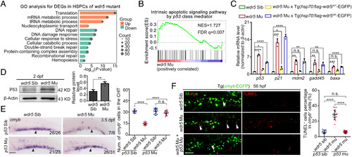
Apoptosis caused by wdr5 deletion is p53-dependent. (A) GO analysis for the downregulated genes in the CD41+gata1− HSPCs of CHT region from wdr5 mutant×Tg(CD41:GFP;gata1:dsRed), compared with that from siblings. (B) GSEA for the upregulated genes associated with the p53-involved signaling pathway in the CD41+gata1− HSPCs of CHT region from wdr5 mutant×Tg(CD41:GFP;gata1:dsRed) compared with that from siblings. (C) qRT-PCR showing the expression of p53, p21, mdm2, gadd45, and baxa in the CHT region of siblings, wdr5 mutants, wdr5 mutant×Tg(hsp70:flag-wdr5WT-EGFP) and wdr5 mutant×Tg(hsp70:flag-wdr5MF-EGFP) at 3.5 dpf. wdr5WT and wdr5MF driven by hsp70 promoter were induced by heat shock at 28 hpf. Error bar, mean ± SD, n.s.: no significance, *P ≤ 0.05, **P ≤ 0.01, ***P ≤ 0.001, ****P ≤ 0.0001. (D) Western blotting (Left) and statistical data (Right) showing the protein level of P53 in the CHT region of siblings and wdr5 mutants at 2 dpf. Error bar, mean ± SD, **P ≤ 0.01. (E) WISH (Left) and statistical data (Right) showing the expression of cmyb (black arrowheads) in the CHT region at 3.5 dpf. Error bar, mean ± SD, n.s.: no significance, ****P ≤ 0.0001. (Scale bar, 100 μm). (F) TUNEL (Left) and statistical data (Right) showing the apoptotic cmyb+ cells (white arrowheads) in the CHT region at 56 hpf. Error bar, mean ± SD, n.s.: no significance, ****P ≤ 0.0001. (Scale bar, 50 μm). |