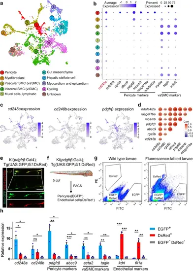
cd248a is highly expressed in pericytes.a Using publicly available single-cell sequencing data set (GSE223922), we show a UMAP projection of 3866 non-skeletal muscle cells collected from whole zebrafish embryos at 50 different developmental stages between 14 and 120 hpf. b Dot plots showing the expression of cd248a, cd248b, and selected pericyte and vascular smooth muscle cell (vaSMC) markers in the indicated clusters. The size of the dot corresponds to the percentage of cluster cells expressing the indicated gene. Color intensity reflects the relative mean expression per cell. c UMAP plots of gene expression (blue dots) highlight individual cells that express cd248a, cd248b, and pdgfrβ. Darker blue dots indicate higher gene expression, while gray dots indicate no gene expression. d Correlation analysis of cd248a, cd248b and other pericyte marker genes. e Labeling of FISH-cd248a (shown in red) and anti-GFP (shown in green) in the Tg(pdgfrβ:Gal4; UAS:GFP) larvae at 4 dpf. Scale bars = 50 μm. f Schematic of the experimental design for sorting pericytes and endothelial cells. g Graphed data of representative FACS analysis of EGFP+ and DsRed+ cells from dissociated 5 dpf Tg(pdgfrβ:Gal4; UAS:GFP; fli1:DsRed) larvae. h qRT-PCR analyses of cd248a, cd248b, pericyte marker genes (pdgfrβ and abcc9), vaSMC marker genes (acta2 and tagln) and endothelial cell marker genes (kdrl and fli1a) followed FACS sorting of EGFP+, DsRed+ and double-negative cells. Data were analyzed by One-way ANOVA and presented as mean ± SD (n = 3 biologically independent experiments). *, p < 0.05. **, p < 0.01. ***, p < 0.001. ns: no significance.
|
