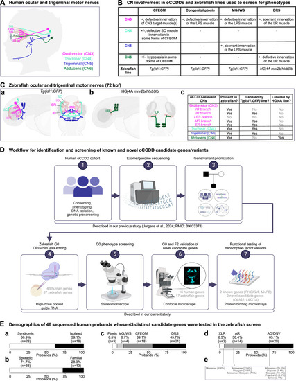
Human and zebrafish cranial motor nerves and study workflow. (A) In the human brainstem, motor neurons cluster into cranial motor nuclei, whose axons form the CNs. The oCCDDs can arise from defective formation, identity, and/or axonal projections of three of these motor neuron populations (CN3, CN4, and CN6) that collectively innervate seven extraocular muscles to orchestrate eye/eyelid movement. CN3 innervates the IO, IR, LPS, MR, and SR muscles. CN4 innervates the SO muscle, and CN6 innervates the LR muscle. CN5 normally innervates the muscles of mastication (not shown) but can also aberrantly innervate the LPS muscle in MGJWS. (B) Summary of CN pathology in humans or mice with specific oCCDDs, and the zebrafish reporter lines used to analyze them. To model oCCDDs in zebrafish, our study leveraged the HGj4A mnr2b/hlxb9lb line for DRS candidate genes and the Tg(isl1:GFP) line for CFEOM, congenital ptosis, and MGJWS genes. (C) Zebrafish wild-type ocular and trigeminal motor nerves visualized in the Tg(isl1:GFP) (a) or HGj4A reporter line (b). Left-lateral view; right-dorsal view. (c) Summary of oCCDD-relevant CNs that are present in zebrafish and labeled by each reporter line. (a, c) As in humans, zebrafish CN3 innervates the IO, IR, MR, and SR muscles. However, the CN3 branch to the IO muscle cannot be visualized with the Tg(isl1:GFP) transgenic line. Additionally, unlike humans, zebrafish lack the LPS muscle and its corresponding CN3 subdivision. Zebrafish have bilateral CN4 motor nuclei and nerves (left and right). CN4 exits the brainstem dorsally and travels ventrally to innervate the contralateral SO muscle. At 72 hpf, wild-type zebrafish CN4 is variably defasciculated. Zebrafish have anterior and posterior trigeminal motor nuclei bilaterally, which extend axons for the motor trigeminal (CN5) nerve that innervates the muscles of mastication (not shown). (b, c) The HGj4A line labels the CN6 motor neurons and their axons. Zebrafish have anterior and posterior CN6 motor nuclei bilaterally, which extend CN6 nerves that target the LR muscles. (D) Workflow for the identification and screening of known and novel oCCDD candidate genes and variants. We reported steps 1-3 in our previous study describing exome/genome sequencing of our human oCCDD cohort3 and performed steps 4–7 in the present study. (E) Demographics of the 46 human probands whose 43 distinct human candidate genes were tested in the zebrafish screen. Percentages of probands are shown with oCCDDs that are (a) syndromic or isolated, (b) sporadic or familial, (c) fitting various oCCDD subdiagnoses, or (d) fitting various modes of inheritance. (e) The types of variants fitting modes of inheritance defined in d are provided. AD, autosomal dominant; AR, autosomal recessive; CFEOM, congenital fibrosis of the extraocular muscles; CN, cranial nerve; CN3, oculomotor/cranial nerve 3; CN4, trochlear/cranial nerve 4; CN5, trigeminal motor/cranial nerve 5; CN6, abducens/cranial nerve 6; DNV, de novo variant; DRS, Duane retraction syndrome; hpf, hours post-fertilization; IO, inferior oblique muscle; IR, inferior rectus muscle; LPS, levator palpebrae superioris muscle; LR, lateral rectus muscle; MGJWS, Marcus Gunn jaw-winking syndrome; MR, medial rectus muscle; oCCDD, ocular congenital cranial dysinnervation disorder; Ptosis, congenital ptosis; SO, superior oblique muscle; SR, superior rectus muscle; XLR, X-linked recessive. Pink, CN3; light green, CN4; dark blue, CN5; dark green, CN6; partially transparent brown, extraocular muscle; dashed line, nerve that is present in zebrafish but not labeled with the transgenic reporter line.
|
