|
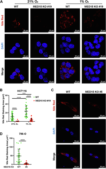
Loss of MED15 suppresses HIF-induced lipid droplet accumulation.A and B, Nile Red and DAPI staining assay of WT and MED15 KO (KO #19) HCT116 cells under normoxia (21% O2) or hypoxia (1% O2) for 72 h. Representative images from three independent experiments are shown in (A). Quantification of Nile Red staining area is shown in (B). Data are represented as means ± SD. n = 27 (21% O2 WT); 25 (21% O2 MED15 KO #19); 26 (1% O2 WT); 30 (1% O2 MED15 KO #19). ∗∗∗p < 0.01, ∗∗∗∗p < 0.0001 by two-way ANOVA with Tukey's multiple comparisons test. C and D, Nile Red and DAPI staining assay of WT and MED15 KO (KO #5) 786-O cells. Representative images from three independent experiments are shown in (C). Quantification of Nile Red staining area is shown in (D). Data are represented as means ± SD. n = 21 (WT); 20 (MED15 KO #5). ∗∗∗∗p < 0.0001 by Student's t test. HIF, hypoxia-inducible factor; med15, Mediator complex subunit 15.
|