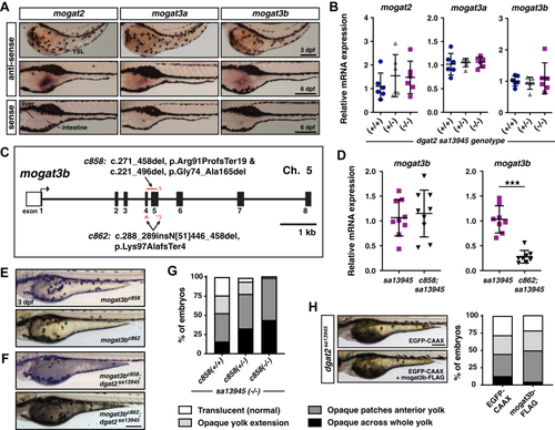
Mogat3b is not responsible for the residual triacylglycerol synthesis in the dgat2 mutants.A, in situ hybridization for mogat2, mogat3a and mogat3b expression at 3 and 6 dpf in wild-type AB embryos. mogat mRNA is not detected in the YSL, but is expressed in the intestine; images are representative of all embryos from three independent experiments at each stage (n = 4–6 embryos per probe per experiment); Scale bars = 200 μm. B, quantitative RT-PCR for mogat2, mogat3a and mogat3b expression in wild-type, dgat2sa13945/+, and dgat2sa13945 embryos at three dpf (N = 6 clutches; 10 pooled fish per sample/genotype, One-way ANOVA, p = 0.8820 mogat1, p = 0.5285 mogat2, p = 0.6988 mogat3b). C, depiction of the mogat3b gene structure highlighting the nature and locations of the c858 and c862 CRISPR/Cas9 mutations (GRCz11; mogat3b ENSDARG00000003635, transcript 201 (ENSDART00000015136.10)). D, quantitative RT-PCR for mogat3b expression in dgat2sa13945versus mogat3bc858;dgat2sa13945 or mogat3bc862;dgat2sa13945 double mutant embryos at 3 dpf (N = 3 independent experiments; 8–9 fish per genotype; unpaired t test, p = 0.6778 c858, ∗∗∗p < 0.001 c862). E, representative images of mogat3bc858 and mogat3bc862 homozygous mutant embryos at 3dpf; Scale = 200 μm. F, representative images of mogat3bc858;dgat2sa13945 and mogat3bc862;dgat3sa13945 double mutant embryos at 3 dpf; Scale = 200 μm. G, embryos from in-crosses of mogat3bc858/+;dgat2sa13945 parents were imaged and scored at 3 dpf for the degree of yolk opacity, binned into the four noted categories prior to genotyping and expressed as a percent of total embryos per genotype (N = 3 independent experiments, n = 62–130 fish per genotype, Chi-square test p < 0.0001). H, dgat2sa13945 embryos were co-injected at the 1-cell stage with CMV:mogat3b-FLAG and CMV:EGFP-CAAX plasmids, or CMV:EGFP-CAAX alone as a control. Bright-field images were obtained of all the embryos that expressed EGFP-CAAX in the YSL at 3 dpf; representative images of embryos from the two treatment groups (left, Scale = 200 μm). Images were assessed for the degree of yolk opacity, binned into the four noted categories of yolk opacity as noted in Fig. 6G and expressed as a percent of total EGFP-positive embryos/treatment group (n = 75 EGFP-CAAX and n = 119 mogat3b-FLAG embryos pooled from three independent experiments, Chi-square test p = 0.0923).
|
