
FIGURE
Fig. 7
- ID
- ZDB-FIG-241219-73
- Publication
- Ranasinghe et al., 2024 - Rotenone exposure causes features of Parkinson`s disease pathology linked with muscle atrophy in developing zebrafish embryo
- Other Figures
- All Figure Page
- Back to All Figure Page
Fig. 7
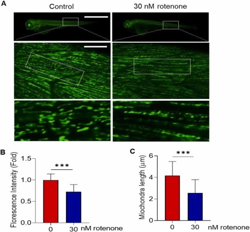
Rotenone exposure induced mitochondria fragmentation in developing zebrafish embryos. Tg(mito:EGFP) embryos aged 7–8 h post fertilization (hpf) were exposed with 30 nM rotenone to 72 hpf. (A) Representative fluorescence (top) and confocal (middle and bottom) microscopic images. Top scale bar: 1 mm, middle scale bar: 10 µm. Quantification of (B) whole body fluorescence intensity and (C) mitochondria length. Ten mitochondria were randomly selected from the dotted box of the middle image of (A), N = 11–12. Data presented as the mean ± SD. SD: standard deviation. * ** p < 0.001, ns: not significant. |
Expression Data
Expression Detail
Antibody Labeling
Phenotype Data
Phenotype Detail
Acknowledgments
This image is the copyrighted work of the attributed author or publisher, and
ZFIN has permission only to display this image to its users.
Additional permissions should be obtained from the applicable author or publisher of the image.
Reprinted from Journal of hazardous materials, 480, Ranasinghe, T., Seo, Y., Park, H.C., Choe, S.K., Cha, S.H., Rotenone exposure causes features of Parkinson`s disease pathology linked with muscle atrophy in developing zebrafish embryo, 136215136215, Copyright (2024) with permission from Elsevier. Full text @ J. Hazard. Mater.