
Fig. 2
- ID
- ZDB-FIG-241119-11
- Publication
- Hoshino et al., 2024 - Pharmacological Inhibition of the Spliceosome SF3b Complex by Pladienolide-B Elicits Craniofacial Developmental Defects in Mouse and Zebrafish
- Other Figures
- All Figure Page
- Back to All Figure Page
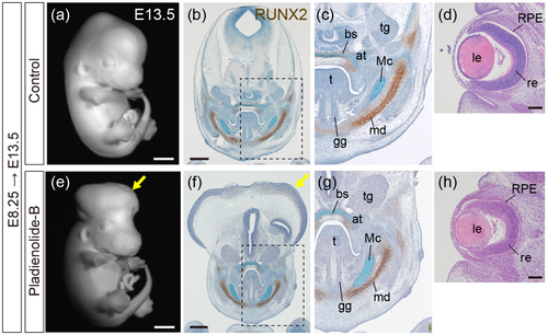
Histological analysis of wild-type fetuses exposed to PB. (a, e) Whole-mount fetuses at E13.5, control (a) and PB-exposed at E8.25 (e). (b, c, f, g) Immunohistochemistry for RUNX2 (brown) with Alcian blue and Toluidine blue staining. Frontal sections of E13.5 control fetuses (b, c) and fetuses exposed to PB at E8.25 (f, g). (c, g) Magnified views of the areas outlined by hatched lines in (b, f). (d, h) Hematoxylin–Eosin staining of frontal sections of the eye at E13.5 in control fetuses (d) and fetuses exposed to PB at E8.25 (h). bs, basisphenoid bone; tg: Trigeminal ganglion; at, ala temporalis; t, tongue; Mc, Meckel cartilage; gg, genioglossus muscle; le, lens; md, mandible; re, retina; RPE, retinal pigment epithelium. Scale bar: 1 mm for (a, e), 500 μm for (b, f), and 100 μm for (d, h). n = 3 obtained from one ICR pregnant mouse, and consistent results were obtained. |