
FIGURE
Fig. EV3
Fig. EV3
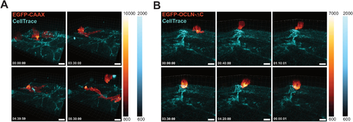
Monocytic OCLN transiently polarizes at monocyte-endothelial interaction sites during transmigration. (A, B) 3D time-lapse spinning disk confocal microscopy of transmigrating monocytes as in Fig. 1E, F. (A) Imaging of a primary monocyte transduced with EGFP-CAAX on hCMEC/D3 monolayer. Images were taken every 10 min. Scale bar: 10 μm. Full video can be found in Movie EV3. (B) Imaging of a primary monocyte transduced with EGFP-OCLN-ΔC on hCMEC/D3 monolayer. Images were taken every 10 min. Scale bar: 10 μm. Full video can be found in Movie EV4. Source data are available online for this figure. |
Expression Data
Expression Detail
Antibody Labeling
Phenotype Data
Phenotype Detail
Acknowledgments
This image is the copyrighted work of the attributed author or publisher, and
ZFIN has permission only to display this image to its users.
Additional permissions should be obtained from the applicable author or publisher of the image.
Full text @ EMBO Rep.