
|
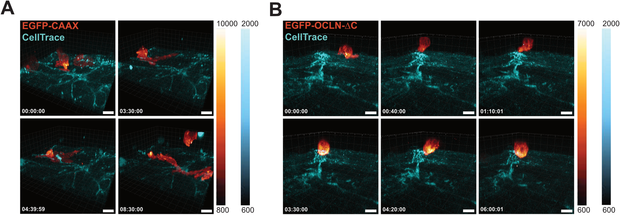
Fig. EV3 Monocytic OCLN transiently polarizes at monocyte-endothelial interaction sites during transmigration. (A, B) 3D time-lapse spinning disk confocal microscopy of transmigrating monocytes as in Fig. 1E, F. (A) Imaging of a primary monocyte transduced with EGFP-CAAX on hCMEC/D3 monolayer. Images were taken every 10 min. Scale bar: 10 μm. Full video can be found in Movie EV3. (B) Imaging of a primary monocyte transduced with EGFP-OCLN-ΔC on hCMEC/D3 monolayer. Images were taken every 10 min. Scale bar: 10 μm. Full video can be found in Movie EV4. Source data are available online for this figure.