
Fig. EV2
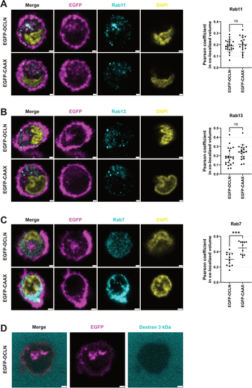
Characterization of the OCLN-containing compartment. (A–C) Immunofluorescence image of primary monocytes expressing either EGFP-OCLN or EGFP-CAAX (magenta) attached to hCMEC/D3 cells and stained with DAPI (yellow) and antibodies (cyan) against Rab11 (A), Rab13 (B), or Rab7 (C). Scale bar: 1 μm, except in C for EGFP-CAAX: 2 μm. (D) Primary monocyte expressing EGFP-OCLN (magenta) attached to hCMEC/D3 monolayer were incubated with 3 kDa fluorescent Dextran (cyan) and immediately imaged using confocal microscopy. The snapshots highlight that Dextran does not access the OCLN-containing compartment. Scale bar: 1 μm. Data information: In (A–C), data are presented as mean ± SEM. Two-tailed Student’s t-test p value < 0.001 (***) or non-significant (ns). Source data are available online for this figure. |