
Fig. EV5
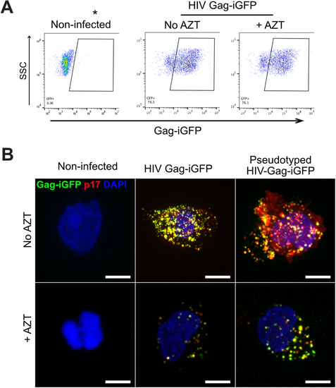
Characterization of primary monocyte infection by HIV-1 and transmigration. (A) Primary monocytes were non-infected or infected with HIV-1 (NLAD8) at MOI 1 for 48 h in the presence or absence of 10 µM AZT. The dot plots show the percentage of Gag-iGFP-expressing cells as a function of the side scatter (SSC) measurement as determined by flow cytometry. The data highlights that despite AZT treatment, monocytes are positive for Gag-iGFP as they carry fluorescent particles. (B) Primary monocytes were non-infected, infected with HIV-1 Gag-iGFP, or HIV-1 Gag-iGFP pseudotyped with VSV-G, at MOI 1 for 48 h in the presence or absence of AZT. Cells were fixed and stained for Gag p17 (red) and DAPI (blue) and Gag-iGFP is shown in green. Scale bar: 5 µm. |