FIGURE

Figure 2.

ID
ZDB-FIG-231005-16
Publication
Anticevic et al., 2023 - Tyrosyl-DNA phosphodiesterase 1 (TDP1) and SPRTN protease repair histone 3 and topoisomerase 1 DNA-protein crosslinks in vivo
Other Figures
All Figure Page
Back to All Figure Page
Figure 2.

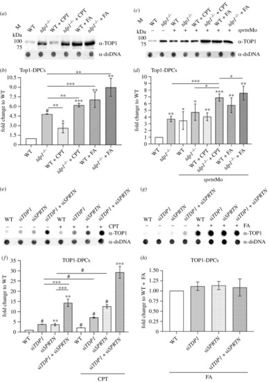
Tdp1 deficiency causes strong accumulation of endogenous and chemically induced Top1-DPCs in embryos and cells. (a) Western blot showing zebrafish Top1-DPCs in tdp1 mutant embryos before and after camptothecin (CPT) (10 µM, 1 h) and formaldehyde (FA) treatment (5 mM, 30 min) (DPC equivalent of 1 µg DNA was loaded per well) and (b) corresponding quantification (n = 4). (c) Western blot showing zebrafish Top1-DPCs in tdp1 mutant embryos before and after sprtn silencing and CPT (10 µM, 1 h) and FA treatment (5 mM, 30 min) and (d) corresponding quantification. (e) Dot blots showing human TOP1-DPCs detected with TOP1-specific antibody before and after CPT treatment of RPE1 cells (50 nM, 1 h) with corresponding DNA loading controls (DPC equivalent of 500 ng DNA was loaded per well). (f) Quantification of (e) from three different biological replicates normalized to untreated WT cells. (g) Dot blots showing human TOP1-DPCs detected with TOP1-specific antibody before and after FA treatment of RPE1 cells (1 mM, 20 min) with corresponding DNA loading controls (DPC equivalent of 500 ng DNA was loaded per well). (h) Quantification of (g) (n = 3). Results represent mean fold change ± s.d. of three different experiments. Statistically significant changes as a result from unpaired Student's t-test are shown as *p < 0.05, **p < 0.01, ***p < 0.001 or #p < 0.0001.

Expression Data

Expression Detail
Antibody Labeling
Phenotype Data

Phenotype Detail
Acknowledgments
This image is the copyrighted work of the attributed author or publisher, and ZFIN has permission only to display this image to its users. Additional permissions should be obtained from the applicable author or publisher of the image. Full text @ Open Biol.