
Fig. 7
- ID
- ZDB-FIG-250612-32
- Publication
- Shi et al., 2025 - Insights into the role of Fsh signaling in ovarian differentiation of chorionic gonadotropin α ( cgα)-deficient zebrafish
- Other Figures
- All Figure Page
- Back to All Figure Page
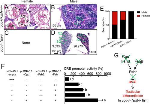
Functional analysis of Fsh signaling function in ovarian differentiation in zebrafish A–D: No ovarian differentiation was observed in cgα−/−;fshr−/− fish at 4 mpf. E: Sex ratio of cgα+/+;fshr+/+ control, fshr−/−, and cgα−/−;fshr−/− fish at 4 mpf. F: cAMP-responsive CRE-luciferase reporter assay in CHO cells transfected with zebrafish Fshr, Fshβ/Fshr, or Cgα/Fshβ/Fshr constructs demonstrated both ligand-independent (constitutive) activity of Fshr and enhanced signaling via Fshβ, with further amplification upon Cgα co-expression. G: Proposed working model of Fsh signaling in ovarian differentiation. SG, spermatogonia; SC, spermatocytes; SZ, spermatozoa; PG, primary growth follicle; PV, previtellogenic follicle; EV, early vitellogenic follicle; MV, middle vitellogenic follicle; FG, full-grown follicle. Letters in bar charts represent significant differences. Green crosses denote gene knockout or inactivation, red letters indicate up-regulation or male-biased sex ratio. CRE, cAMP response element. |