
Fig. 4
- ID
- ZDB-FIG-250612-29
- Publication
- Shi et al., 2025 - Insights into the role of Fsh signaling in ovarian differentiation of chorionic gonadotropin α ( cgα)-deficient zebrafish
- Other Figures
- All Figure Page
- Back to All Figure Page
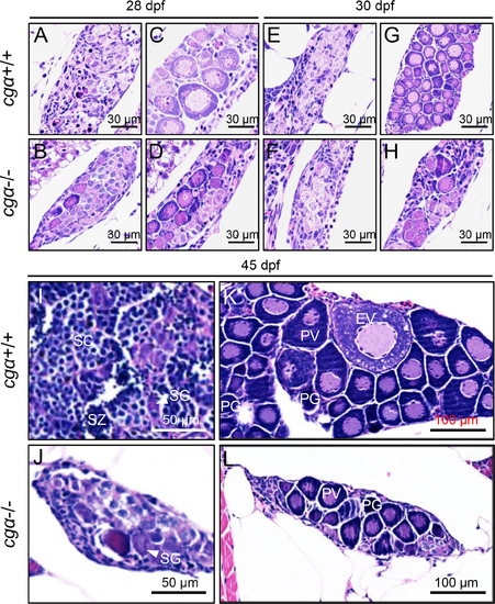
Histological analysis of gonadal development in control and cgα-deficient fish during key stages of sex differentiation A–H: No obvious gonadal differences were observed between control and cgα-deficient fish at 28 and 30 dpf. I, J: At 45 dpf, testes from cgα-deficient males were visibly smaller than those of control males, with germ cells predominantly at the spermatogonia stage. K, L: At 45 dpf, ovaries of cgα-deficient females were smaller than those of control females and contained only PV and PG oocytes. dpf, days post-fertilization; SG, spermatogonia; SC, spermatocytes; SZ, spermatozoa; PG, primary growth follicle; PV, previtellogenic follicle; EV, early vitellogenic follicle. |