
Fig. 5
- ID
- ZDB-FIG-250430-23
- Publication
- Zi et al., 2024 - Protocol for generating a pericyte reporter zebrafish line Ki(pdgfrb-P2A-GAL4-VP16) using a CRISPR-Cas9-mediated knockin technique
- Other Figures
- All Figure Page
- Back to All Figure Page
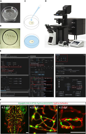
In vivo imaging of zebrafish brains using a confocal microscope (A) Photograph of a 60-mm plastic dish with a 14-mm glass coverslip-filled well. (B) Photographs the larvae embedded in the glass coverslip-filled well of a 60-mm plastic dish. (C) Schematic showing the operation that add ES or Hank’s solution (marked as blue) for the embedded larvae before imaging. (D) Photograph of a FV3000 confocal microscope (Olympus) for in vivo imaging of zebrafish. (E) Screenshot of the software interface (FV31S-SW) for operating the FV3000. The red box marks the settings for scan speed, resolution, aperture, laser intensity, detector voltage, z-series, and z-step positions. (F) Representative images of pericytes and blood vessels in the brain of Ki(pdgfrb-P2A-Gal4-VP16);Tg(4×nrUAS:GFP);Tg(fli1a:DsRed) larvae. These images are processed with the software ImageJ. Pericyte in green, endothelial cell in red. Scale bar 50 μm. |