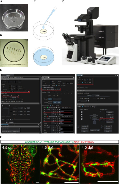
Fig. 5 In vivo imaging of zebrafish brains using a confocal microscope (A) Photograph of a 60-mm plastic dish with a 14-mm glass coverslip-filled well. (B) Photographs the larvae embedded in the glass coverslip-filled well of a 60-mm plastic dish. (C) Schematic showing the operation that add ES or Hank’s solution (marked as blue) for the embedded larvae before imaging. (D) Photograph of a FV3000 confocal microscope (Olympus) for in vivo imaging of zebrafish. (E) Screenshot of the software interface (FV31S-SW) for operating the FV3000. The red box marks the settings for scan speed, resolution, aperture, laser intensity, detector voltage, z-series, and z-step positions. (F) Representative images of pericytes and blood vessels in the brain of Ki(pdgfrb-P2A-Gal4-VP16);Tg(4×nrUAS:GFP);Tg(fli1a:DsRed) larvae. These images are processed with the software ImageJ. Pericyte in green, endothelial cell in red. Scale bar 50 μm.
Image
Figure Caption
Acknowledgments
This image is the copyrighted work of the attributed author or publisher, and
ZFIN has permission only to display this image to its users.
Additional permissions should be obtained from the applicable author or publisher of the image.
Full text @ STAR Protoc

