Figure 4
- ID
- ZDB-FIG-250328-22
- Publication
- Jia et al., 2025 - FRET-Based Sensor Zebrafish Reveal Muscle Cells Do Not Undergo Apoptosis in Starvation or Natural Aging-Induced Muscle Atrophy
- Other Figures
- All Figure Page
- Back to All Figure Page
|
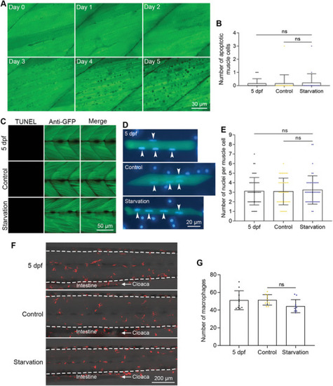
Muscle cells did not undergo apoptosis during starvation‐induced muscle atrophy. A) FRET imaging of muscle cells during zebrafish starvation. The time points after starvation are indicated in each image. B) The quantified results show the number of apoptotic muscle cells during starvation (n = 20 zebrafish for each group). C) TUNEL assays show that no muscle cells die of apoptosis after starvation. D) Nuclear staining of dissociated muscle cells. Nuclei from muscle cells are indicated with arrowheads. E) The quantified results show the number of nuclei of muscle cells after starvation‐induced muscle atrophy (n = 45 cells for each group). F) Live imaging of macrophages in the trunk regions of zebrafish after starvation‐induced atrophy. The intestine and cloaca are marked to show the images were taken from the same region. G) The quantified results show the number of macrophages after starvation‐induced muscle atrophy (n = 10 zebrafish for each group). The size of each scale bar is indicated in each image. ns: no significant difference. |

