
Figure 2
- ID
- ZDB-FIG-250328-20
- Publication
- Jia et al., 2025 - FRET-Based Sensor Zebrafish Reveal Muscle Cells Do Not Undergo Apoptosis in Starvation or Natural Aging-Induced Muscle Atrophy
- Other Figures
- All Figure Page
- Back to All Figure Page
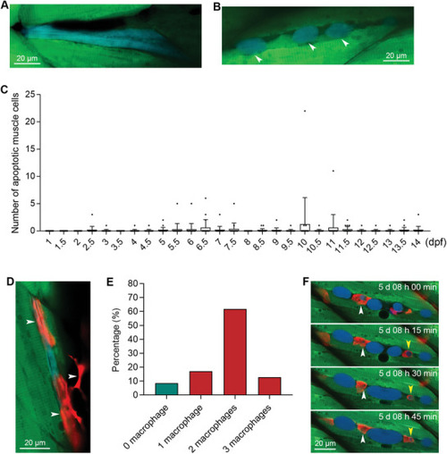
Sensor zebrafish can be used for visualizing developmental muscle cell apoptosis and the engulfment of dead muscle cells by multiple macrophages. A,B) Apoptotic muscle cells during normal zebrafish development. The blue muscle cell in was degraded into smaller fragments, indicated by arrowheads. C) The quantified results show the number of apoptotic muscle cells per zebrafish at different time points during normal development (n = 20 zebrafish for each time point). D) The engulfment of an apoptotic muscle cell by macrophages. The macrophages are indicated with white arrowheads. E) Percentages of apoptotic muscle cells that colocalized with single or multiple macrophages. F) Live tracking of the engulfment of a dead muscle cell by red macrophages. The developmental time is indicated in each image. Two apoptotic bodies engulfed by macrophages are indicated with white and yellow arrowheads, respectively. The size of each scale bar is indicated in each image. |