
Fig. 1 - Supplemental 3
- ID
- ZDB-FIG-250327-51
- Publication
- Kondrychyn et al., 2025 - Combined forces of hydrostatic pressure and actin polymerization drive endothelial tip cell migration and sprouting angiogenesis
- Other Figures
-
- Fig. 1
- Fig. 1 - Supplemental 1
- Fig. 1 - Supplemental 2
- Fig. 1 - Supplemental 3
- Fig. 2
- Fig. 3
- Fig. 3 - Supplemental 1
- Fig. 3 - Supplemental 2
- Fig. 3 - Supplemental 3
- Fig. 3 - Supplemental 4
- Fig. 3 - Supplemental 5
- Fig. 3 - Supplemental 6
- Fig. 3 - Supplemental 7
- Fig. 4
- Fig. 4 - Supplemental 1
- Fig. 5
- Fig. 6
- Fig. 7
- All Figure Page
- Back to All Figure Page
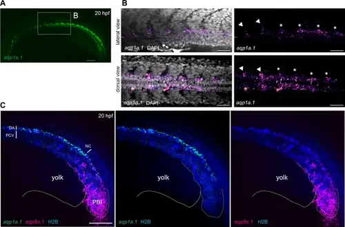
aqp1a.1 and aqp8a.1 are differentially expressed in the dorsal aorta at 20 hpf. (A, B) aqp1a.1 mRNA expression in the DA, as determined by RNAscope in situ hybridization. (B) Magnified region of the anterior trunk. aqp1a.1 expression is heterogeneous, with higher expression in endothelial tip cells of newly formed intersegmental vessels (ISVs) (arrowhead) and in endothelial cells (ECs) of the DA destined to be tip cells (asterisk). (C) aqp8a.1 mRNA expression in the posterior blood island, as determined by RNAscope in situ hybridization. Endothelial nuclei are labeled with EGFP (pseudocolored in blue) in Tg(fli1a:H2B-EGFP)nvc69 transgenic line. Representative confocal images of nine embryos (n = 2 independent experiments). Anterior to the left. DA, dorsal aorta; NC, notochord; PCV, posterior cardinal vein; PBI, posterior blood island. Scale bar, 100 µm (A) and 40 µm (B, C). |