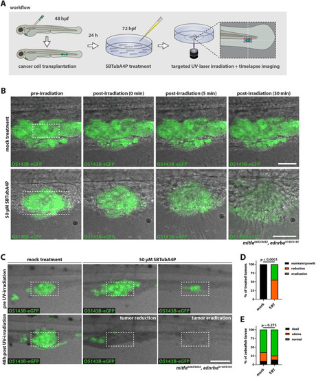
Targeted SBTubA4P illumination eliminates metastases in vivo. (A) Schematic representation of the workflow for SBTubA4P-mediated ablation of disseminated tumor cells in a zebrafish xenograft model. Transplantation of cancer cells into the perivitelline space was performed at 2 dpf. After 24 h [1 day post injection (dpi)], larvae were selected for disseminated tumor cells in the caudal hematopoietic tissue (CHT), treated with 50 µM SBTubA4P for 4 h, anesthetized, washed and mounted for spatially targeted UV irradiation. Mock-treated controls were not treated with SBTubA4P. Single microtumors in the CHT were targeted by repeated UV scanning of a rectangular ROI over the course of 10 min. (B) SBTubA4P illumination induces rapid cell death in xenografted tumor cells. OS143B-xenotransplanted zebrafish larvae at 1 dpi were treated as outlined in A. The subsequent timelapse after UV illumination reveals induction of spatially confined tissue damage at the targeted area, with loss of eGFP expression and extrusion of cells over the course of 30 min in SBTubA4P-treated larvae (see Movie 7). Dashed line rectangles indicate the targeted ROI (70×35 µm). Representative confocal images from two independent experiments (n=3-4 per condition). Scale bars: 50 µm. (C) SBTubA4P illumination ablates OS143B xenograft microtumors in vivo (see Movie 8). Zebrafish larvae at 1 dpi were treated as outlined in A, then removed from the imaging dishes, washed and kept until 5 dpf (48 h post treatment) for follow-up imaging. Dashed line rectangles indicate areas targeted by UV irradiation (140×70 µm). Representative confocal images (maximum projections: ten planes, 5 µm spacing) from three independent experiments. Scale bar: 100 µm. (D) Quantification of SBTubA4P-mediated OS143B tumor treatment efficacy. Chi-squared test was performed for comparison of mock and SBTubA4P (SBT) treatments on outcome: maintenance or growth, reduction and total eradication of tumor masses. Stacked bars represent fractions of total larvae from three independent experiments (mock, n=21; SBT, n=18). (E) Quantification of SBTubA4P treatment toxicity following xenograft tumor treatment (OS143B and SK-N-MC cells). Chi-squared test was performed for comparison of mock and SBTubA4P treatment toxicity, scoring for occurrence of dead larvae, edema formation or no adverse effects. Stacked bars represent fractions of total larvae from five independent experiments (mock, n=40; SBT, n=43).
|
