|
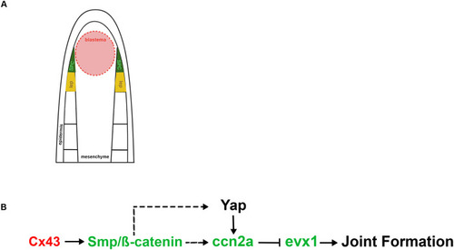
Proposed model for interactions of ccn2a within the Cx43-dependent joint formation pathway. (A) Schematic representation of a longitudinal section of a zebrafish fin ray. The blastema (red), located distally and medially, is composed of proliferative cells driving regeneration. Skeletal precursor cells (SPCs, green), situated laterally, will differentiate into either osteoblasts or joint-forming cells. Newly formed lepidotrichia, or the skeletal elements, are shown in yellow. (B) Cx43 (acting in the cells of the medial blastema, denoted by red text) promotes Smp/β-catenin (acting in the SPCs, denoted by green text), which may promote ccn2a expression (also in SPCs). We show that Ccn2a inhibits evx1, which is required for joint formation. Yap may be regulated downstream of β-catenin or independently.
|