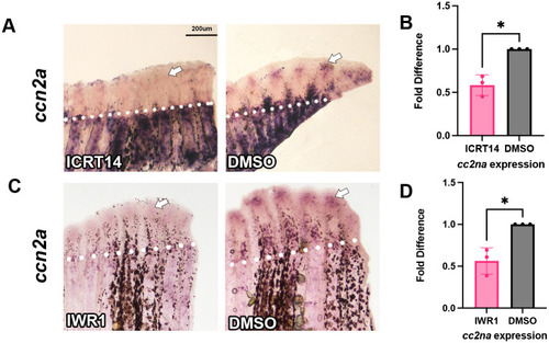
ccn2a expression was decreased with β-catenin inhibition. Fins were amputated at 50% and treated with either 5 µM ICRT14 via injection or 10 µM IWR1 introduced into the water at 72 hpa. Fins were harvested 24 h later at 96 hpa. Amputation planes are denoted by white dotted line. In situ hybridization was performed using antisense digoxygenin-labeled probe against ccn2a to measure relative gene expression. (A) There was a decrease in ccn2a expression in ICRT14 treated fins compared to DMSO. (B) Reduction of gene expression was quantified through qPCR in ICRT14 treated fins. Graph shows a mean of three biological replicates fold difference and standard deviation (n=5 fins per replicate). A fold difference of 1 means no change from wild-type expression. The Student's t-test (two tailed, unpaired) was used to assess significance. (C) There was a decrease in ccn2a expression in IWR1 treated fins compared to DMSO. (D) Reduction of gene expression was quantified through qPCR in IWR1 treated fins. Graph shows a mean of three biological replicates fold difference and standard deviation (n=5 fins per replicate). A fold difference of 1 means no change from wild-type expression. The Student's t-test (two tailed, unpaired) was used to assess significance. Scale bar: 200 µm.
|
