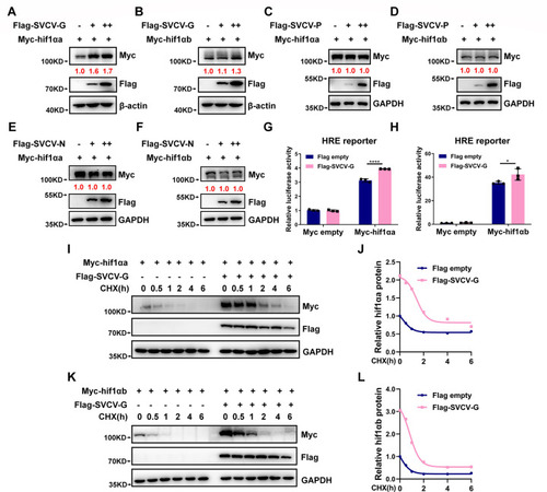
SVCV-G protein stabilizes hif1αa and hif1αb. (A, B) Immunoblotting (IB) of Myc-hif1αa (A) and Myc-hif1αb (B) in HEK293T cells transfected with the plasmid expressing Myc-hif1αa or Myc-hif1αb together with increasing amounts of Flag-SVCV-G (Flag empty vector [-] was used as a control). (C, D) IB of Myc-hif1αa (C) and Myc-hif1αb (D) in HEK293T cells transfected with the plasmid expressing Myc-hif1αa or Myc-hif1αb together with increasing amounts of Flag-SVCV-P (Flag empty vector [-] was used as a control). (E, F) IB of Myc-hif1αa (E) and Myc-hif1αb (F) in HEK293T cells transfected with the plasmid expressing Myc-hif1αa or Myc-hif1αb together with increasing amounts of Flag-SVCV-N (Flag empty vector [-] was used as a control). (G, H) HRE reporter activity in HEK293T cells transfected with the plasmid expressing Myc-hif1αa (G) or Myc-hif1αb (H) together with Flag-SVCV-G (Flag empty vector was used as a control). (I, J) IB of the indicated proteins in HEK293T cells transfected with Myc-hif1αa together with Flag-SVCV-G for 24 h (Flag empty vector [-] was used as a control) and then treated with cycloheximide (CHX, 50 µg/mL) for an increasing time (0–6 h) (I). The relative intensities of Myc-hif1αa were determined by normalizing the intensities of Myc-hif1αa to the intensities of GAPDH (J). (K, L) IB of the indicated proteins in 293T cells transfected with Myc-hif1αb together with Flag-SVCV-G for 24 h (Flag empty vector [-] was used as a control) and then treated with CHX (50 µg/mL) for an increasing time (0–6 h) (K). The relative intensities of Myc-hif1αb were determined by normalizing the intensities of Myc-hif1αb to the intensities of GAPDH (L).
|
