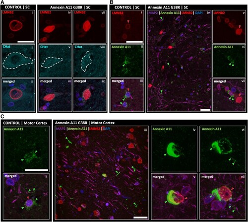
Immunostaining of Lamin B2 and Annexin A11 in controls and Annexin A11 mutant ALS+/FTD cases. (A) Immunostaining of LMNB2 in a representative control and spinal cord (SC) of anterior horn neurons from the G38R case. Tissue was stained for LMNB2, ChAT (choline acetyltransferase) to label motor neurons and DAPI. A representative ChAT positive motor neuron [A(ii)] displaying a nuclear envelope localized LMNB2 signal [A(ii and iii)]. Two ChAT positive motor neurons [A(v and viii)] from the G38R case showing abnormal nucleoplasmic LMNB2 [A(iv, vi and vii, ix), respectively]. Images presented as 3D z-stack projections. Scale bar = 50 µm. (B) Immunostaining of a representative control and spinal cord of anterior horn neurons from the G38R case displaying the difference in Annexin signal. Wild-type ring-like LMNB2 [B(i), red arrow] and cytoplasmic diffuse Annexin signal [B(ii), green arrow] in a representative control neuron [B(iii)]. Images presented as 3D z-stack projections. Single plane image from the anterior horn region of spinal cord tissue from the G38R case [B(iv)]. Neurite aggregates of Annexin can be seen (green arrows). Representative image of neurons reiterating the LMNB2 nucleoplasmic signal [B(vi and vii, red arrow)] and displaying a more punctate Annexin signal [B(vi and vii, green arrows)]. Images presented as 3D z-stack projections. Scale bar = 50 µm. (C) Immunostaining of a representative control and motor cortex neurons from the G38R case showing a marked presence of Annexin aggregation. Representative control motor cortex neuron displaying a diffuse cytoplasmic Annexin signal [C(i and ii)]. Motor cortex from the G38R case showing nucleoplasimc LMNB2 signal and neuropil Annexin aggregates (green arrows) [C(iii)]. Neurons from the G38R case motor cortex reporting heavy perinuclear Annexin aggregates [C(iv, v and vi, vii), respectively; green arrows for Annexin aggregates, red arrows for LMNB2 nucleoplasmic signal]. Images presented as 3D z-stack projections. Scale bar = 50 µm.
|
