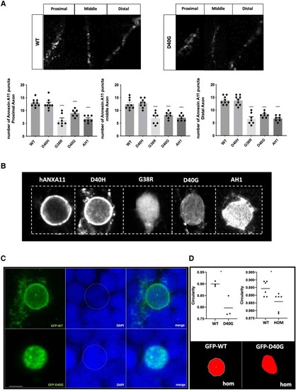
WT-Annexin A11 is present as puncta throughout the axon and is reduced due to ALS or AH1 del mutations. (A) Representative single plane images of wild-type (WT) and D40G Annexin A11 positive puncta in the proximal, middle and distal axon segment (top). Puncta were quantified in axons from larvae injected with controls WT Annexin A11, D40H and mutants D40G, G38R and AH1 del. Total n caudal primary (CaP) neurons = 39 (one per embryos): WT-Annexin A11-eGFP (n = 9), D40H-Annexin A11-eGFP (n = 9), G38R-Annexin A11-eGFP (n = 7), D40G-Annexin A11-eGFP (n = 7) and AH1-Annexin A11-eGFP (n = 7) (bottom). All three mutations reduce the amount of Annexin A11 puncta in each segment. ****P ≤ 0.0001, one-way ANOVA, Tukey’s multiple comparison test. (B) Annexin A11 mutations alter Annexin A11 nuclear envelope distribution. Qualitative GFP immunofluorescence shown left to right, wild-type, D40H, G38R, D40G and AH1del-Annexin A11-eGFP and counterstained with DAPI (signal not shown). Images are ×63 and ×3 digital zoom (Zeiss LSM 800). (C) Misexpression of human WT Annexin A11 can rescue nuclear envelope integrity in homozygous Annexin A11a knockout larvae. Top (left to right): Wild-type Annexin A11-eGFP when expressing in knockout Annexin A11a larvae localizes to the nuclear envelope and as perinuclear puncta (eGFP and DAPI merge). Bottom (left to right): D40G Annexin A11-eGFP by contrast is completely nucleoplasmic with the presence of intra-nuclear aggregates (eGFP and DAPI merge). Scale bar = 5 μm. (D) Nuclear circularity changes are associated with loss of Annexin A11a or the D40G mutation in Annexin A11. Bottom: Representative images from circularity analysis of homozygous knockout larvae expressing wild-type or D40G Annexin A11-GFP. Quantification (top left) shows a significant deviation from circularity. *P ≤ 0.05, unpaired t-test wild-type (n = 3) and D40G (n = 3). Similarly, F3 homozygous knockout larvae (top right) show a deviation of nuclear circularity from F3 wild-type larvae of the same line, unpaired t-test *P ≤ 0.05 [WT (n = 6) and homozygous (Hom, n = 6)]. ALS = amyotrophic lateral sclerosis; GFP = green fluorescent protein.
|
