
FIGURE
Figure 5
- ID
- ZDB-FIG-231225-186
- Publication
- Huysseune et al., 2023 - Bone Formation in Zebrafish: The Significance of DAF-FM DA Staining for Nitric Oxide Detection
- Other Figures
- All Figure Page
- Back to All Figure Page
Figure 5
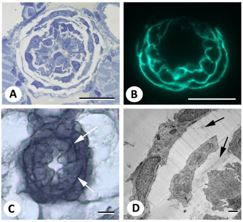
Demonstration of elastic fibers in the bulbus arteriosus. ( |
Expression Data
Expression Detail
Antibody Labeling
Phenotype Data
Phenotype Detail
Acknowledgments
This image is the copyrighted work of the attributed author or publisher, and
ZFIN has permission only to display this image to its users.
Additional permissions should be obtained from the applicable author or publisher of the image.
Full text @ Biomolecules