
Fig. 3
- ID
- ZDB-FIG-211122-3
- Publication
- Duan et al., 2021 - The unique structure of the zebrafish TNF-α homotrimer
- Other Figures
- All Figure Page
- Back to All Figure Page
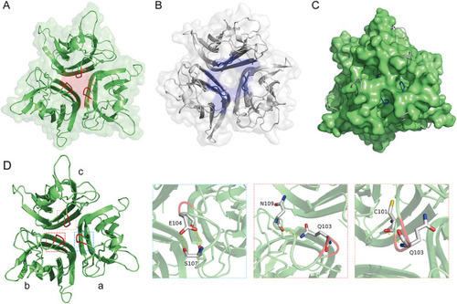
The unique topological structure of the zTNF-α1 trimer. (A) Top view of the trimeric zTNF-α1 structure colored green. The part of the EF loop at the top that folds inward into the trimer channel is colored red. (B) Top view of the trimeric hTNF-α structure colored light gray. The part of the EF loop at the top is colored blue. (C) Superposition of the structure of zTNF-α1 (surface) with the structure of hTNF-α (cartoon). The EF loops of hTNF-α protrude outward at the three corresponding holes formed around the top of the zTNF-α1 trimer. (D) The interaction between residues 103–104 in the EF loop (colored in red) and adjacent amino acids. Key residues forming hydrogen bonds are shown as sticks. Hydrogen bonds are indicated with a yellow dashed line. The corresponding residues are labeled with amino acid abbreviations and primary sequence numbers. |