
Fig. 2
- ID
- ZDB-FIG-211122-2
- Publication
- Duan et al., 2021 - The unique structure of the zebrafish TNF-α homotrimer
- Other Figures
- All Figure Page
- Back to All Figure Page
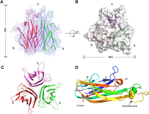
Structural characteristics of the zTNF-α1 trimer. (A) Overview of the electron density map (2Fo - Fc) at 2.6 Å (contoured at 1.5σ) with the refined model superimposed. The threefold axis lies vertically. Double-sided arrows indicate the height of the complex. (B) Top view of the structural surface of trimeric zTNF-α1. Double-sided arrows indicate the width of the complex. (C) A top view of the trimeric zTNF-α1 structure is shown in the cartoon representation. (D) Monomeric zTNF-α1 is colored blue to red from the N-terminus to the C-terminus, as shown in the cartoon representation. Secondary structure elements and disulfides are labeled. |