
Figure 3.
- ID
- ZDB-FIG-210518-66
- Publication
- Pham et al., 2020 - HDAC6 promotes growth, migration/invasion, and self-renewal of rhabdomyosarcoma
- Other Figures
- All Figure Page
- Back to All Figure Page
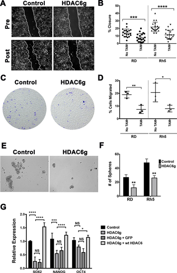
(A) Representative images from a wound healing scratch assay in RD cells with tamoxifen-induced CRISPR/Cas9 HDAC6 targeting. Post = 16 hours post-scratch. Dashed lines indicate migrating fronts. (B) Summary of scratch assays in RD and Rh5 with tamoxifen-inducible CRISPR/Cas9-mediated HDAC6 targeting in RD and Rh5. Each datapoint represents a distinct area of the gap. The results shown are from one representative experiment of at least 3 repeats. (C) Representative images from a transwell migration assay. Cells migrated to the bottom chamber were stained with crystal violet at 22 hours post-seeding in the top chamber. (D) Summary of transwell migration assays in RD and Rh5 with tamoxifen-inducible CRISPR HDAC6 targeting in RD and Rh5. Each data point represents a replicate well. Results shown are from one of 3 independent experiments. (E) Representative images from a sphere assay in RD cells. (F) Summary of sphere assays in RD and Rh5 cell lines 3 days post-plating. Shown are results of 4 replicates from one of 3 independent experiments. (G) RT-PCR analysis comparing expression levels of stem cell markers in RD sphere cells harboring no TAM control and TAM-induced HDAC6-targeting, TAM-induced HDAC6-targeting transduced with GFP expression construct and TAM-induced HDAC6-targeting transduced with Cas9-resistant wild-type HDAC6 expression construct harvested after 3 days of culturing in stem cell medium. Results were the average of 3 replicates from one of 3 independent experiments. Each error bar in B, D, F and G represents standard deviation. Two-tailed t-test in B, D and F; two-way ANOVA test in G; * = p <0.05; ** = p < 0.01; *** = p < 0.001; **** = p < 0.0001. |