
- Title
-
Physical constraints on the positions and dimensions of the zebrafish swim bladder by surrounding bones
- Authors
- Satoh, K., Maeno, A., Adachi, U., Ishizaka, M., Yamada, K., Koita, R., Nakazawa, H., Oikawa, S., Fujii, R., Furudate, H., Kawamura, A.
- Source
- Full text @ J. Anat.

The os suspensorium is more in contact with the anterior end of the swim bladder than the tripus in adult zebrafish. (a) A micro-CT scan image demonstrates the relationship between the swim bladder (red) and the surrounding skeletal structures (white) in wild-type adult zebrafish (n = 4). Movies S1 and S2 provide 3D representations of this relationship. Additionally, a 2D movie, showing the outline of the swim bladder (Movie S3, sagittal) is available. (b–e) Plane sections obtained through micro-CT scanning. The yellow arrowhead indicates the os suspensorium, which directly attaches to the anterior margin of the swim bladder (sb). The white arrowhead represents the posterior edge of the tripus, which is closely associated with the swim bladder. The anterior orientation is to the left. A 2D movie, illustrating these sections is provided as Movie S4. (f, g) The morphology of the os suspensorium is shown. The os suspensorium is an extending bone located medially from the 4th centrum. The portion of the os suspensorium in contact with the swim bladder is demarcated by a bracket. The transverse process of vertebra 4 (tp4), which extends laterally and ventrally from the 4th centrum, is also indicated. A 3D movie illustrating these structures is available as Movie S5. Scale bars: 1000 μm (a), 500 μm (b–e), 300 μm (f, g). |

Anterior protrusion of the swim bladder in hoxc6a mutant fish showing the absence of the os suspensorium and the malformed tripus. (a–f) Representative micro-CT scan images of the anterior vertebrae in wild-type (n = 4) and hoxc6a homozygous adult fish (n = 4). In the images, the os suspensorium is indicated by the yellow arrowhead, while the tripus is indicated by the white arrowhead in wild-type fish. A 3D movie is available in Movie S6. (g) Visualization of the swim bladder (depicted in red) and the surrounding bones in hoxc6a mutant fish (n = 4). The presumptive anterior edge of the swim bladder in normal adult fish is indicated by the yellow dashed line. The 3D movies are available with Movies S7 and S8. Furthermore, a 2D movie illustrating the outline of the swim bladder is provided in Movie S9 (sagittal view). Scale bars: 1000 μm. |
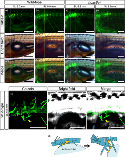
Pushback of the anterior lobe of the swim bladder by the developing os suspensorium. (a–d) The anterior edge of the inflating swim bladder relative to the vertebrae was compared between wild-type (n = 6) and hoxc6a mutants (n = 5). Calcein staining was used to visualize the bones (a-d), which were then merged with the bright field images (a’-d’) to create the combined images (a’’-d’’). The anterior edge of the anterior lobe in the swim bladder is highlighted with a white dashed line. The presumptive position (4th centrum) corresponding to the anterior edge of the swim bladder in typical normal adult zebrafish is indicated by a yellow dashed line. The swim bladder is outlined by a blue dashed line. The number of vertebrae starts from the most anterior vertebra. Images were taken using a fluorescent stereomicroscope from the lateral side. (e) After the calcein staining, the anterior vertebrae and swim bladder of wild-type larvae were visualized using a confocal microscope (n = 3). An oblique dorsoventral view is presented, and the arrowhead indicates the developing os suspensorium attaching to the swim bladder. (f) Schematics illustrate the model showing the counterclockwise rotation of the os suspensorium (depicted in yellow) pushing back the anterior swim bladder (light blue) during zebrafish development. Scale bars: 300 μm. |
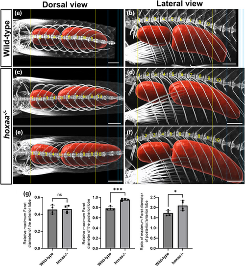
Posterior enlargement of the swim bladder in hoxaa cluster-deleted mutants with an additional pair of pleural ribs. (a–f) Micro-CT scan images showing the swim bladder (red) and the surrounding skeletal structures in wild-type (n = 4) and hoxaa cluster-deleted adult fish (n = 4). To ensure reproducibility, two representative images are shown for hoxaa cluster-deleted adult fish. The vertebrae are numbered in order starting from the first rib-bearing vertebra. To facilitate comparisons, the dimensions of the fish body were standardized based on the distance between the first and ninth rib-bearing vertebra, shown by the yellow dashed line. The blue dashed line indicates the posterior extent of the swim bladder in wild-type fish, while the light blue dashed lines represent the posterior extent of the swim bladder in hoxaa cluster-deficient mutants. A 3D movie is available as Movie S11. (g) A comparison of the maximum Feret diameter of the anterior and posterior lobes between wild-type and hoxaa cluster-deficient mutants. A Student's t-test was performed, with significance indicated as *p < 0.05 and ***p < 0.001. Scale bars: 1000 μm. |