
Fig. 3
- ID
- ZDB-FIG-250417-75
- Publication
- Satoh et al., 2024 - Physical constraints on the positions and dimensions of the zebrafish swim bladder by surrounding bones
- Other Figures
- All Figure Page
- Back to All Figure Page
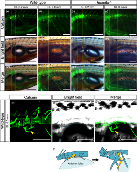
Pushback of the anterior lobe of the swim bladder by the developing os suspensorium. (a–d) The anterior edge of the inflating swim bladder relative to the vertebrae was compared between wild-type (n = 6) and hoxc6a mutants (n = 5). Calcein staining was used to visualize the bones (a-d), which were then merged with the bright field images (a’-d’) to create the combined images (a’’-d’’). The anterior edge of the anterior lobe in the swim bladder is highlighted with a white dashed line. The presumptive position (4th centrum) corresponding to the anterior edge of the swim bladder in typical normal adult zebrafish is indicated by a yellow dashed line. The swim bladder is outlined by a blue dashed line. The number of vertebrae starts from the most anterior vertebra. Images were taken using a fluorescent stereomicroscope from the lateral side. (e) After the calcein staining, the anterior vertebrae and swim bladder of wild-type larvae were visualized using a confocal microscope (n = 3). An oblique dorsoventral view is presented, and the arrowhead indicates the developing os suspensorium attaching to the swim bladder. (f) Schematics illustrate the model showing the counterclockwise rotation of the os suspensorium (depicted in yellow) pushing back the anterior swim bladder (light blue) during zebrafish development. Scale bars: 300 μm. |