
|
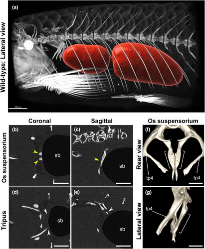
Fig. 1 The os suspensorium is more in contact with the anterior end of the swim bladder than the tripus in adult zebrafish. (a) A micro-CT scan image demonstrates the relationship between the swim bladder (red) and the surrounding skeletal structures (white) in wild-type adult zebrafish (n = 4). Movies S1 and S2 provide 3D representations of this relationship. Additionally, a 2D movie, showing the outline of the swim bladder (Movie S3, sagittal) is available. (b–e) Plane sections obtained through micro-CT scanning. The yellow arrowhead indicates the os suspensorium, which directly attaches to the anterior margin of the swim bladder (sb). The white arrowhead represents the posterior edge of the tripus, which is closely associated with the swim bladder. The anterior orientation is to the left. A 2D movie, illustrating these sections is provided as Movie S4. (f, g) The morphology of the os suspensorium is shown. The os suspensorium is an extending bone located medially from the 4th centrum. The portion of the os suspensorium in contact with the swim bladder is demarcated by a bracket. The transverse process of vertebra 4 (tp4), which extends laterally and ventrally from the 4th centrum, is also indicated. A 3D movie illustrating these structures is available as Movie S5. Scale bars: 1000 μm (a), 500 μm (b–e), 300 μm (f, g).