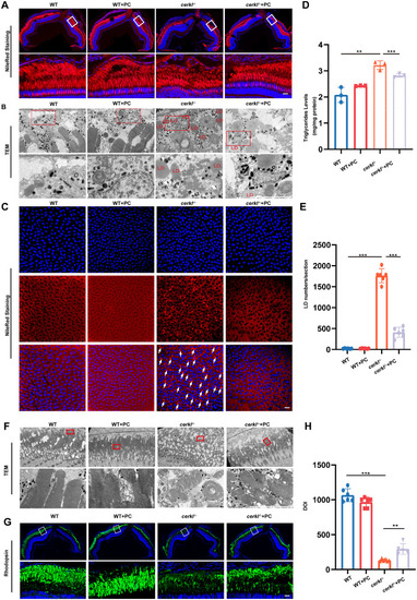
PC addition alleviates LD accumulation and photoreceptor dysfunction in cerkl−/− zebrafish. (A) Confocal imaging of LDs in WT and cerkl−/− retina with PC treatment zebrafish by Nile Red staining. Blue represents DAPI and red represents Nile Red staining. RPE, retinal pigment epithelium; ONL, outer nuclear layer; INL, inner nuclear layer. Scale bars = 10 µm. (B) TEM analysis of LDs in WT and cerkl−/− retina with PC treatment zebrafish. LD, lipid droplets. Scale bars = 10 µm. (C) Confocal imaging of LDs in WT and cerkl−/− RPE with PC treatment zebrafish by Nile Red staining. Blue represents DAPI and red represents Nile Red staining. Scale bars = 10 µm. (D) TG levels in WT and cerkl−/− retina with PC treatment zebrafish by TGs analysis kits. **, P < 0.01, ***, P < 0.001. (E) Quantitative analysis of LDs is shown in D. ***, P < 0.001. (F) TEM analysis of photoreceptors in WT and cerkl−/− retina with PC treatment zebrafish. Bars = 10 µm. (G) Immunofluorescence analysis of retinas with PC treatment zebrafish using anti-Rhodopsin antibody. Scale bars = 10 µm. (H) Quantitative analysis of the rhodopsin is shown in G. **, P < 0.01, ***, P < 0.001.
|
