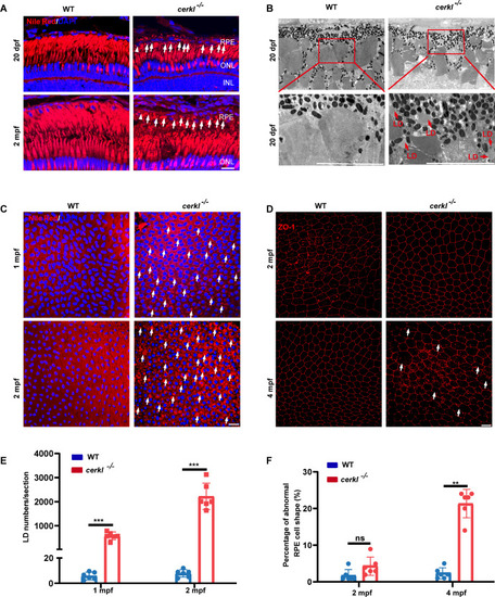
CERKL ablation triggers progressive lipid droplet pathology in RPE. (A) Confocal imaging of LDs in WT and cerkl−/− retina at 20 dpf and 2 mpf zebrafish by Nile Red staining. Blue represents DAPI and red represents Nile Red staining. RPE, retinal pigment epithelium; ONL, outer nuclear layer; INL, inner nuclear layer; Scale bars = 10 µm. (B) TEM analysis of LDs in WT and cerkl−/− zebrafish retina at 20 dpf. LD, lipid droplet. Scale bars = 10 µm. (C) Confocal imaging of LDs in WT and cerkl−/− RPE at 1 mpf and 2 mpf zebrafish by Nile Red staining. Blue represents DAPI and red represents Nile Red staining. Scale bars = 10 µm. (D) Immunofluorescence analysis of RPE cells from WT and cerkl−/− zebrafish at 2 mpf and 4 mpf using anti-ZO-1 antibody. Scale bars = 10 µm. (E) The quantitative analysis of LD numbers shown in C. The results are shown as mean ± SEM. ***, P < 0.001. (F) The numbers of abnormal RPE cells were qualified for each of the images in D. The results are shown as mean ± SEM. **, P < 0.01.
|
