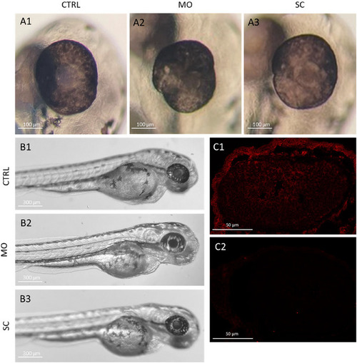
Morpholino oligo‐mediated knockdown does not influence gross eye size and morphology or overall embryo development but does eliminate Rs1 protein production at 72 hpf. A: Gross eye morphology and size at 72 hpf were imaged in the sagittal plane of a representative uninjected control (CTRL) (A1), translation‐blocking morpholino oligo (MO)‐injected (A2), and scrambled control oligo (SC)‐injected (A3) embryos under a dissection microscope. Some variations in eye shape were observed in MO‐ and SC‐injected larvae (A2, A3), but no microphthalmia. The scale bars represent 100 µM. B: At 72 hpf, there were no readily discernible differences in the developmental stage between uninjected (B1) MO‐injected (B2) and SC‐injected (B3) siblings. Deformations associated with oligo overdose were very rare at the chosen oligo concentrations (50 ng/µL; less than 1% in 100 injections). C. Rs1 protein presence was observed at 72 hpf in SC larvae (C1) but not in MO‐injected larvae (C2) using IHC. The scale bars represent 50 µm. WT = wild‐type, MO = morpholino‐oligo injected, SC = control injected, hpf = hours post‐fertilization.
|
