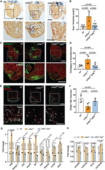
Partial restoration of cardiac regeneration in nr4a1 mutant through reduction of inflammatory response. (A) AFOG staining performed on cardiac sections of the indicated genotypes at 60 dpci. (B) Quantification of scar size in AFOG-stained cardiac sections. (C) Collagen deposition in the hearts of indicated genotypes at 21 dpci, as detected by staining with collagen I antibody. (D) Quantification of collagen deposition in the wounded area at 21 dpci. (E) PCNA/Nkx2.5 double immunostaining to detect proliferating cardiomyocytes in 7 dpci hearts of indicated genotypes. Arrowheads indicate the proliferating cardiomyocytes. (F) Quantification of colocalization of PCNA and Nkx2.5 per unit area in different groups. (G) qPCR of inflammation-related genes in the hearts of indicated genotypes at 7 dpci. (H) qPCR of fibrosis-related genes in the hearts of indicated genotypes at 7 dpci. Dashed lines show the wounded area. Boxed regions in A and E show the position of magnified images at the bottom. Boxed regions in C show the approximate positions for quantification. The fold changes in G and H are calculated relative to the expression in uninjured WT hearts. P-values<0.05 were considered to be statistically significant (two-tailed unpaired t-test in B, D and F; two-way ANOVA with Sidak test for multiple comparison correction and two-tailed unpaired t-test in G and H). Data are mean±s.e.m. Scale bars: 275 µm (A,E); 250 µm (C).
|
