|
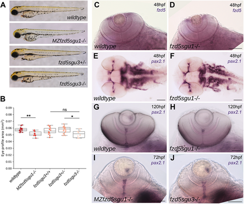
Subtle eye defects in fzd5sgu1, MZfzd5sgu1 and fzd5sgu3 embryos. (A) MZfzd5sgu1 and fzd5sgu3 3 days post-fertilisation (dpf) larvae morphology compared to that of wild-type and heterozygote siblings. (B) Quantifications of projected eye area in the genotypic groups from A. Box plots represent the median and 25-75th percentiles; whiskers indicate the range of the data. Each data point represents one eye. A pairwise Tukey HSD post-hoc test revealed statistically significant eye size differences between the wild-type and the MZfzd5sgu1 group (P=0.006748), and between the fzd5sgu3 homozygote and heterozygote group (P=0.01591). ns, not significant; *P<0.05, **P<0.01. (C-J) Expression of fzd5 in the CMZ (C,D) and pax2.1 in the optic stalk/optic nerve (E-J) in fzd5sgu1 (D,F,H), MZfzd5sgu1 (I) and fzd5sgu3 (J) embryos compared to wild-type controls (C,E,G). Embryo age is indicated in each panel. Scale bars: 100 µm.
|