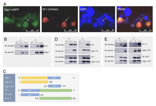
Interaction of Zglp-1 and Sf-1 occurs between their zinc finger domains. A Co-localization of Zglp-1 and Sf-1 was observed in the nucleus of HEK293T cells. Confocal microscopy images taken 24 h after co-transfection revealed the expression of Zglp-1 (depicted in green) and Sf-1 (shown in red) within the cells. Nuclei were counterstained with DAPI (blue). B An interaction analysis between Zglp-1 and Sf-1 was performed through co-immunoprecipitation and immunoblot assays in HEK293T cells co-transfected with Zglp-1-HA (2 μg) and Sf-1-Myc (2 μg). C Domain structure of zebrafish Zglp-1 and Sf-1 predicted by the SMART program. The amino acid sequence of Zglp-1 was divided into two sections. Amino acids 227–374 were designated Zglp-1-ZnF. Amino acids 1–226 were designated Zglp-1-N. The Sf-1 amino acid sequence was segmented into two distinct regions: amino acids 1–163, designated as Sf-1-ZnF, and amino acids 164–502, constituting the Sf-1-C segment. D Interaction studies were conducted in HEK293T cells co-transfected with combinations of Zglp-1-ZnF-HA (2 μg) and Sf-1-Myc (2 μg), or Zglp-1-N-HA (2 μg) and Sf-1-Myc (2 μg). These interactions were analyzed using co-immunoprecipitation and immunoblot techniques. E Similar interaction analyses were performed in HEK293T cells co-transfected with Sf-1-ZnF-Myc (2 μg) and Zglp-1-ZnF-HA (2 μg), or Sf-1-C-Myc (2 μg) and Zglp-1-ZnF-HA (2 μg). Co-immunoprecipitation and immunoblotting were employed to assess the interactions between these proteins
|
