
Fig. 6
- ID
- ZDB-FIG-250521-11
- Publication
- Xu et al., 2025 - Microglia-Derived IL-6 Promotes Müller Glia Reprogramming and Proliferation in Zebrafish Retina Regeneration
- Other Figures
- All Figure Page
- Back to All Figure Page
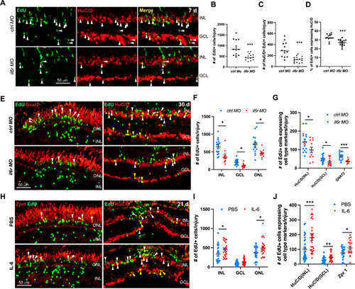
IL-6 signaling promotes the regeneration of retinal neurons in injured retinas. (A) EdU and HuC/D immunofluorescence showing the early MGPC differentiation at 7 dpi in retinas electroporated with 1 mM of control MO (ctrl MO) or il6r MO. (B, C, D) Quantification of the total number of EdU+ cells per injury B, the number of EdU and HuC/D double positive cells per injury C, and the proportion of EdU+ cells expressing HuC/D per injury D in A. (E) Immunofluorescence showing the regeneration of retinal neurons at 30 dpi in retinas electroporated with 1 mM of ctrl MO or il6r MO. (F) Quantification of the number of EdU+ cells in each layer per injury at 30 dpi in E. (G) Quantification of the number of regenerated photoreceptor (ONL Gnat+/EdU+), amacrine cells (INL HuC/D+/EdU+), and RGCs (GCL HuC/D+/EdU+) per injury at 30 dpi in E. (H) Immunofluorescence showing the regeneration of retinal neurons at 21 dpi in retinas treated with PBS control or IL-6. (I) Quantification of the number of EdU+ cells in each layer per injury at 21 dpi in H. (J) Quantification of the number of regenerated photoreceptor (ONL Zpr1+/EdU+), amacrine cells (INL HuC/D+/EdU+), and RGCs (GCL HuC/D+/EdU+) per injury at 21 dpi in H. *, P < 0.05; **, P < 0.01; ***, P <0.001; GCL, ganglion cell layer; INL, inner nuclear layer; ONL, outer nuclear layer. |