
Fig. 3
- ID
- ZDB-FIG-250417-24
- Publication
- Xu et al., 2024 - Single-cell RNA sequencing reveals the heterogeneity and interactions of immune cells and Müller glia during zebrafish retina regeneration
- Other Figures
- All Figure Page
- Back to All Figure Page
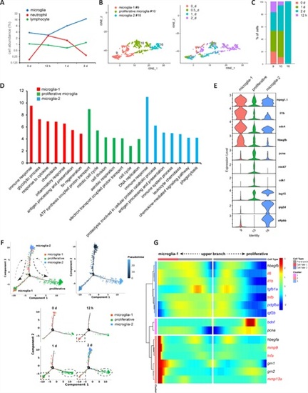
Characterization of microglial heterogeneity and state changes in intact and stab-injured zebrafish retinas.(A) Abundance of different immune cell types in the retinal samples at each time point tested. (B) Clustering of microglia subtypes. (C) Cell population composition in terms of the three microglial subtypes. (D) GO analysis showing the enriched biological processes in the microglia subtypes. (E) Subtype-specific marker genes. (F) Pseudotime analysis of microglial subtypes. (G) BEAM analysis of secreted factors during microglial state transition. Red, pro-inflammatory factors; blue, anti-inflammatory factors. BEAM: Branched Expression Analysis Modeling; GO: Gene Ontology. |